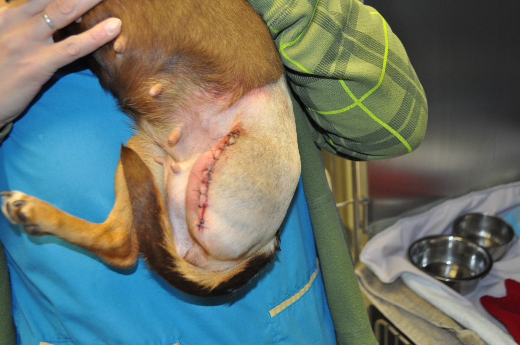
Fully narrated surgical videos.
In depth guide and more.
Pick the appropriate level of amputation:
• A mid femoral amputation is appropriate for most hind limb conditions:
-Fractures of the proximal third femur and distal.
-Neoplasms that are at/or distal to the stifle (tumour dependent).
• A disarticulation amputation is appropriate for:
-Fractures of the femoral neck or coxofemoral joint
-Neoplasms located in the mid to distal femur or the stifle (tumour dependent).
• A hemi/semi pelvectomy is appropriate for:
-Neoplasms in the proximal femur including the coxofemoral joint (tumour dependent).
-If the patient is using the limb well (perhaps the amputation is for an aggressive subcutaneous soft tissue sarcoma), consider placing them in an Ehmer sling for 3-4 days prior to the surgery so that they can learn to ambulate on 3 limbs prior to the surgery.
•Dogs and cats can get around quite well with a hind limb amputation; even the large breeds.
• They may want to make changes to their home to accommodate their pet, especially in the early days as the pet is adjusting:
-Have non-slippery surfaces for the pet to ambulate on.
-Lower the bed/couch/favorite chair by removing the legs (on the furniture, of course).
-Have a harness to assist the pet in the first days (especially the bigger dogs) when handling stairs.
-Consider unplugging the doorbell if the dog reacts excitedly when it rings (at least for the early days).
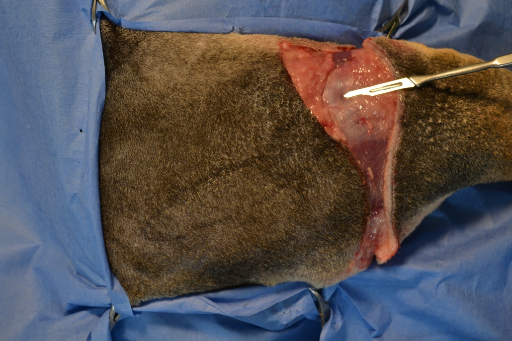
- Figure 1
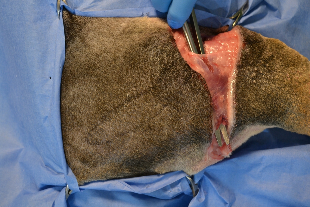
- Figure 2
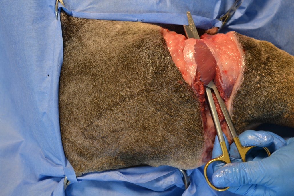
- Figure 3
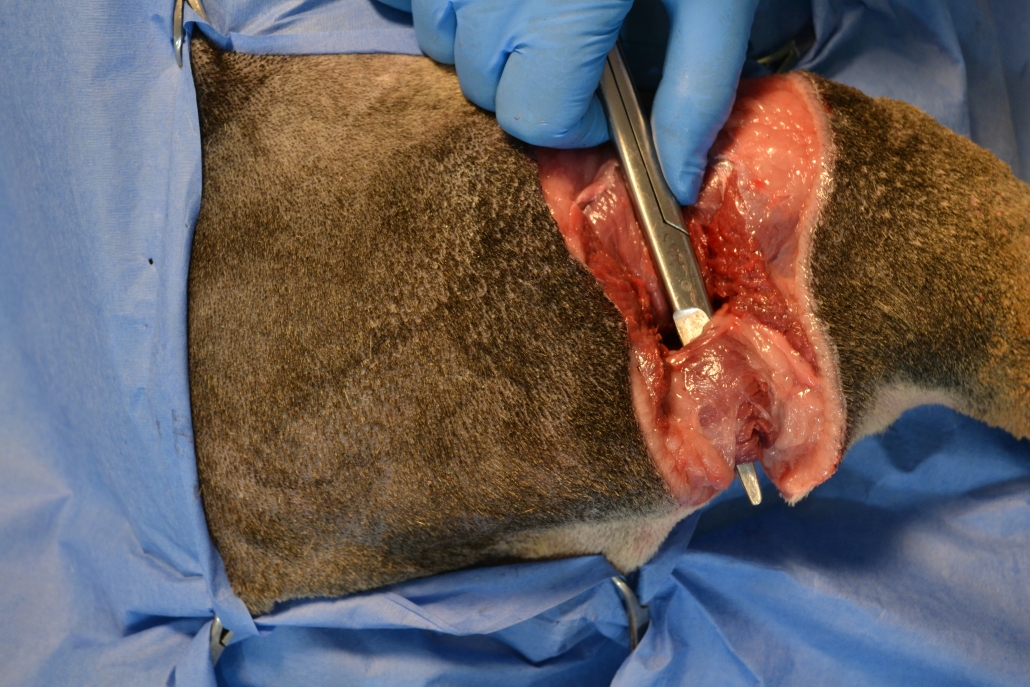
- Figure 4
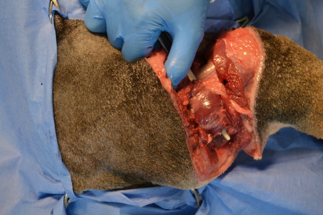
- Figure 5
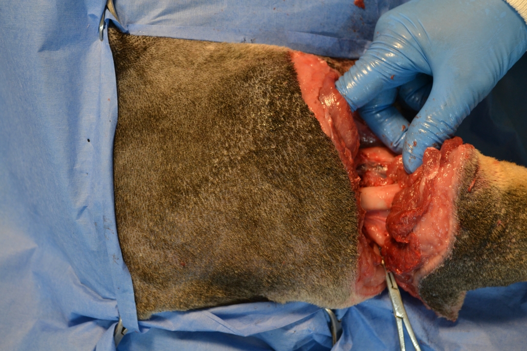
- Figure 6
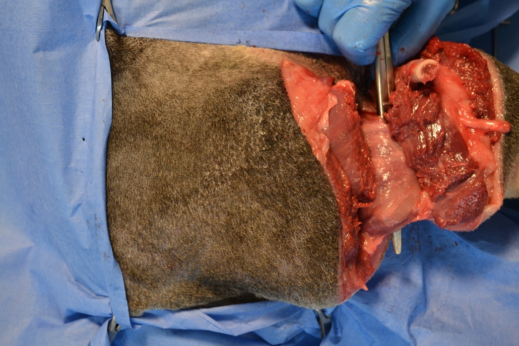
- Figure 7
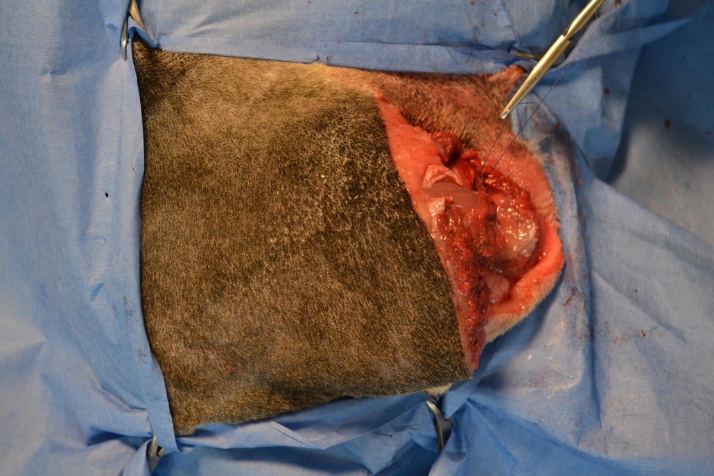
- Figure 8
• Have an abundance of small mosquito hemostats.
• The larger the patient, the more hemostats you should have.
• A patellar saw, hobby saw or a gigli wire can be used to cut the femur.
• Drains are rarely necessary with this procedure.
• Manage their pain, especially for the first 24-48 hours. It diminishes rapidly after that time frame.
• Help with support (belly towel or chest harness) for the first few walks.
• Have non-slippery surfaces for them move on especially in the early days.
• Use antibiotics as indicated by the conditions and/or length of the procedure.
The best way to be more confident when doing this procedure is to have an experienced surgeon looking over your shoulder guiding you and encouraging you.
The information in this virtual workshop is the next best thing. Surgery is more than following a step-by-step pictorial description. It is important to be comfortable with the anatomy of the area. We will help you understand the relevant anatomy from a surgical perspective.
There are fully narrated videos; (1) is a narrated presentation that explains when and how to do the surgery, how to select your cases and managing the patient post-operatively. There are narrated videos of mid-femoral amputations performed on a cadaver, so it is easy to see the details; and on a patient, so you can appreciate how it really looks. There are also videos on the disarticulation surgery as well.
If you do plan on doing this surgery; have an anatomy book close at hand! When you watch the video, listen carefully to my words, I give a lot of little details that will help make the surgery easier for you. You will see how to handle the instruments and the tissues. Watch/listen to the video several times so that when you are in surgery you will continue to hear my voice.
Subscribe to our newsletter and receive 15% off a workshop of your choice!
Workshops & Courses
Blog
 The Secret to having the Bandage stay Above the StifleJuly 3, 2024 - 1:44 pm
 Common Mistakes made by Veterinarians when doing the Drawer TestJune 26, 2024 - 9:22 am
 Burying the end knot in a continuous subcutaneous suture patternJune 19, 2024 - 7:35 pm
 Creating Donuts for your BandagesJune 12, 2024 - 3:15 pm
 The Knotless Suture PatternJune 5, 2024 - 2:45 pm

